
Study finds iron-rich enamel protects, but doesn’t color, rodents’ orange-brown incisors
 strengthen and protect the teeth. Credit: Adapted from ACS Nano 2024, DOI: 10.1021/acsnano.4c00578")
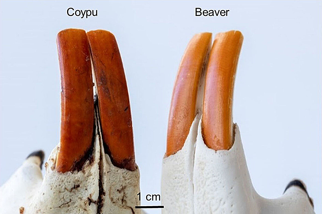
Chattering squirrels, charming coypus, and tail-slapping beavers—along with some other rodents—have orange-brown front teeth. Researchers have published high-resolution images of rodent incisors in ACS Nano, providing an atomic-level view of the teeth’s ingenious enamel and its coating. They discovered tiny pockets of iron-rich materials in the enamel that form a protective shield for the teeth but, importantly, don’t contribute to the orange-brown hue—new insights that could improve human dentistry.
Human and animal teeth are coated in a complex crystalline substance called enamel. And while enamel is the hardest tissue in our bodies, it’s even harder in rodents. Their ever-growing incisors have an additional outer layer of acid-resistant, iron-rich enamel.
Previously, researchers suggested that this iron-rich material was also responsible for the striking orange to brown color of many rodents’ incisors. However, the microscopic structure of the iron-rich enamel hadn’t been fully characterized. To learn more about the composition of rodent tooth enamel, Vesna Srot and colleagues captured high-resolution images of incisor specimens from several species.
The researchers collected incisors from rodents that live in different environments: beavers, coypus, squirrels, marmots, rats, voles and mice. To investigate the structure, elemental composition and color transmission of the enamel, thin slices were taken from different sections of the teeth and prepared for imaging with optical microscopy, 3D focused ion beam tomography and scanning transmission electron microscopy. The micro- and nano-scale resolution images revealed:
- Initially, cells that synthesize enamel components produce 6- to 8-nanometer-wide particles of iron-storage proteins called ferritins, which are the source material for iron ions in matured enamel.
- As enamel matures and solidifies before the teeth erupt from the gums, iron-containing ferrihydrite-like material moves into the outer layer of enamel, occupying empty spaces between calcium-containing hydroxyapatite crystals.
- The microstructure of the iron-rich enamel contains elongated nanometer-sized pockets filled with small amounts of the ferrihydrite-like material, which contribute acid resistance even though the filled pockets account for less than 2% of the volume of iron-rich enamel.
- While these results suggest that different types of rodents develop the iron-rich outer enamel layer in a similar way, the depth of the layer vary by species, with mice having the thinnest and coypus having the thickest layers.
- Finally, the intense orange-brown color of rodent incisors doesn’t come from the filled pockets in the enamel, as was previously thought, but from a thin surface layer composed of aromatic amino acids and inorganic minerals.
The researchers suggest that adding small amounts of ferrihydrite-like or other colorless biocompatible iron minerals to dental care products could provide exceptional protection for human tooth enamel. In addition, incorporating small amounts of iron hydroxides into synthetic enamel could produce longer-lasting restorations for human teeth.
More information:
Ingenious Architecture and Coloration Generation in Enamel of Rodent Teeth, ACS Nano (2024). DOI: 10.1021/acsnano.4c00578
Provided by
American Chemical Society
Citation:
Study finds iron-rich enamel protects, but doesn’t color, rodents’ orange-brown incisors (2024, April 17)
retrieved 17 April 2024
from https://phys.org/news/2024-04-iron-rich-enamel-doesnt-rodents.html
This document is subject to copyright. Apart from any fair dealing for the purpose of private study or research, no
part may be reproduced without the written permission. The content is provided for information purposes only.